

Increased blood flow, indicating a possible infectionĪre you looking for an affordable venous ultrasound? At North Atlanta Vascular and Vein Center (Suwanee, Lawrenceville, Roswell and Cumming), we provide safe, accurate, and affordable ultrasounds.Decreased or missing blood flow to any organ.Tumors and inborn vascular malformations.This can be observed in congenital vascular abnormalities and dialysis fistula.ĭoppler ultrasound images can help doctors evaluate: Review a link between an artery and a vein.In children, venous ultrasound is used to: Checking a blood vessel graft used for dialysis.Helping to guide the positioning of a needle or catheter in a vein.Determine the cause of long-standing leg swelling.

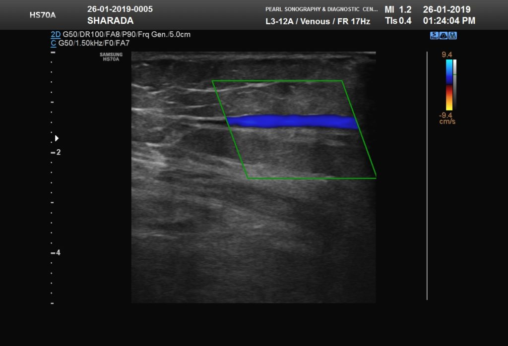
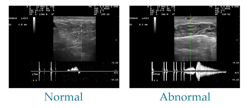
A venous ultrasound examination can include a Doppler ultrasound study. Venous ultrasound gives images of the veins all through the human body. They can reveal the internal organs of the body's structure and movement as well as the blood flowing into vessels of the blood. High-frequency sound waves pass through the body, and photographs are taken in real-time. Venous ultrasound imagingĭuring ultrasounds, a tiny probe called a transducer and gel are mounted directly on the skin. They are also known as thrombosis of the deep vein, or DVT. Searching for blood clots, especially in the leg’s veins, is the most common reason for a venous ultrasound test. It is widely used, especially in the veins of the leg, to check for blood clots. In order to create images of the veins in the body, venous ultrasound uses sound waves.


 0 kommentar(er)
